With full compensatory pause, next normal beat arrives an interval is equal double preceding R-R interval. Retrograde capture describes process the ectopic impulse conducted retrogradely the AV node, producing atrial depolarisation. is visible the ECG an inverted P wave ("retrograde P wave"), occurring the QRS complex.
 A observational analysis Kim al published 2021 the to suggest the risk new‐onset AF higher patients PVCs. 5 this issue the Journal the American Heart Association (JAHA), Lee co‐authors describe new association specifically PVC burden new‐onset AF, irrespective underlying diseases, a single‐center .
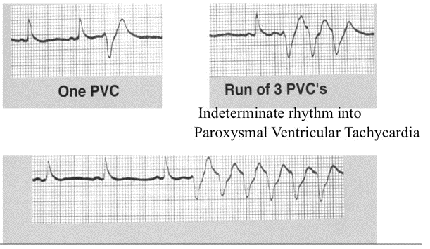
 The morphology the PVC vary the ECG Rhythm more one area the ventricles irritable spontaneously discharge, designated multifocal mulitiform PVCs. suggests more irritable ventricle sometimes lead the rhythm Ventricular Tachycardia. occurrence may trigger .

 The fact salvos AF triggered PVCs continued occur the procedure failed sustain suggests the left atrial surface area conduction sufficiently diminished PVI preclude sustained AF, without directly targeted clinical AF trigger. fact, PVC ablation may treated patient .
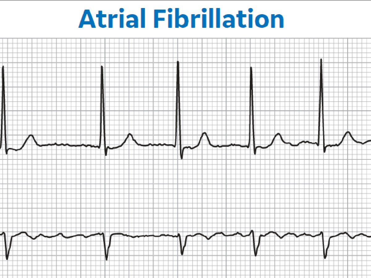
 An ECG with paroxysmal AF PVC simultaneously detected the top panel Fig. 5. AF detected a sliding window the totally irregular RR intervals disappearing P. addition, PVC beat also recognized having regularly irregular RR interval, widened QRS duration, less cross-correlations, is measure .
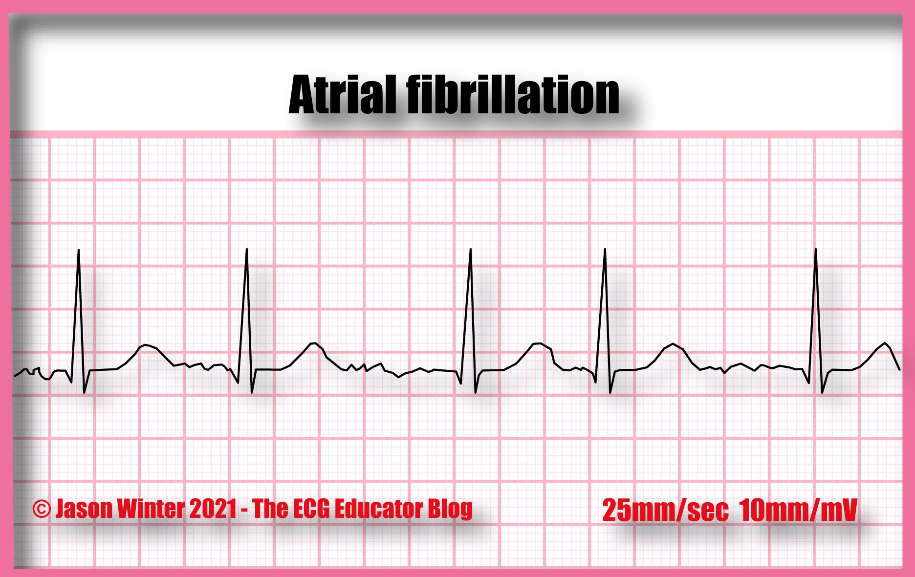 An A-Fib.com reader me email about difference Atrial Fibrillation PVCs. start, PVC stands Premature Ventricular Contraction. is PVC?… Premature Ventricular Contraction (PVC) like extra beat a missed beat comes the part your heart, ventricles. to worry.
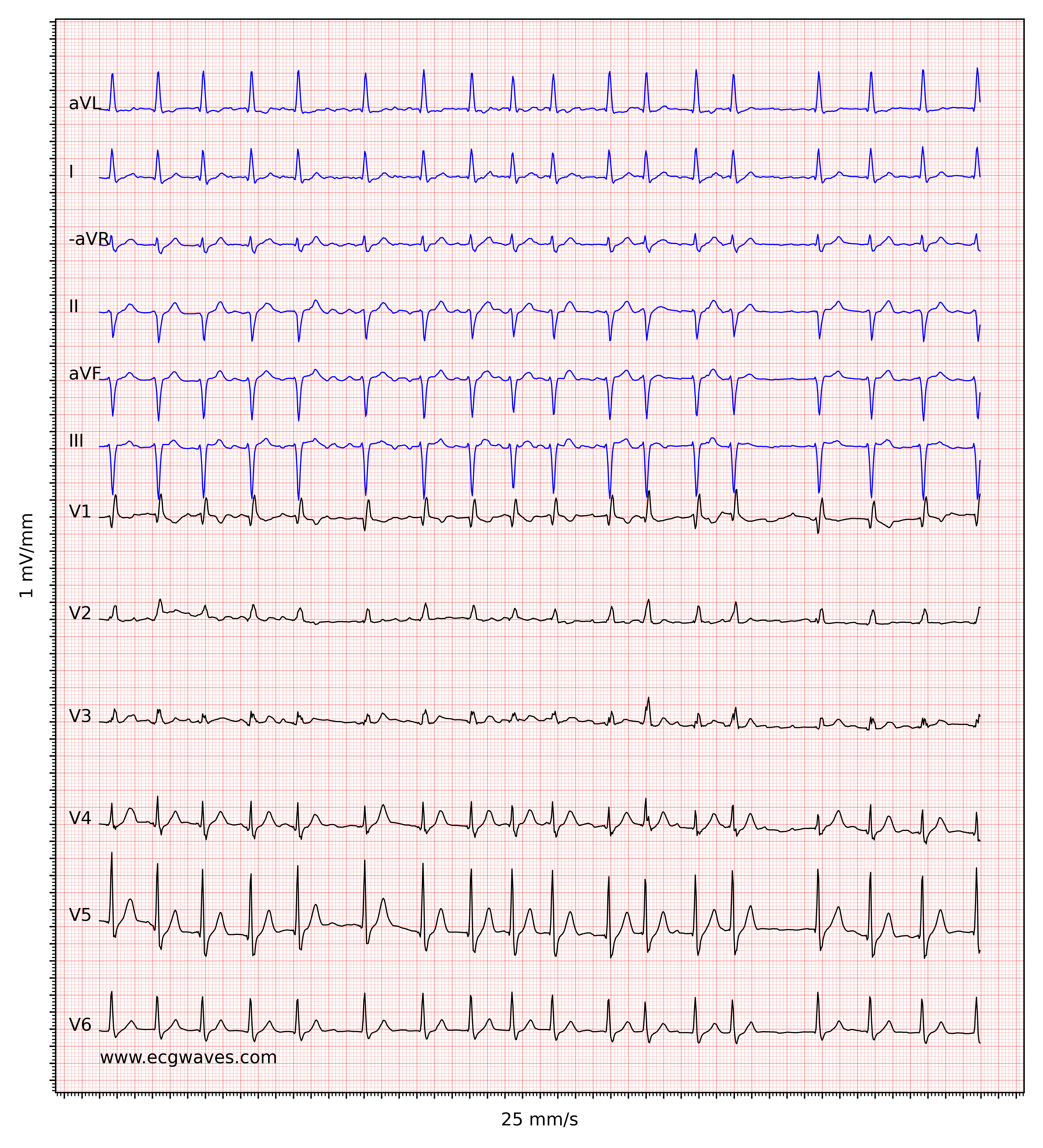 As PVCs infrequent most patients, brief period an electrocardiogram fail capture ectopic beats. also the differentiation a PVC ectopic atrial beats, are termed premature atrial contractions (PACs). patients PVCs, ECG reveal findings include:
 In normal subjects, isolated ventricular extrasystoles found about 1% subjects subjected standard ECG. healthy subjects undergoing dynamic ECG recording 24-48h, extrasystoles be frequent (> 60 PVC hour), monomorphic (i.e. a single morphology), rarely polymorphic (in 5% cases) (of multiple .
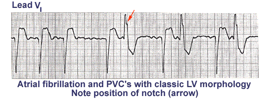
 The QRS morphology EKG predict PVCs site origin. a broad general rule, right ventricular ectopic pacemaker generates ventricular complex left bundle branch block (LBBB) pattern, the left ventricular ectopic pacemaker generates ventricular complex right bundle branch block (RBBB) pattern 2. right left ventricular outflow tracts aortic cusp .
 ECG Learning Center - An introduction to clinical electrocardiography
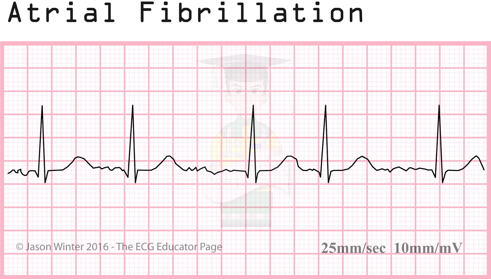 ECG Educator Blog : Atrial Rhythms
 Visual interpretation of the model a, b The ECGs of AF and PVCs, which
 What Premature Ventricular Contraction (PVC) Looks Like on Your Watch
 VT AF PVCs | ECG-Cafe
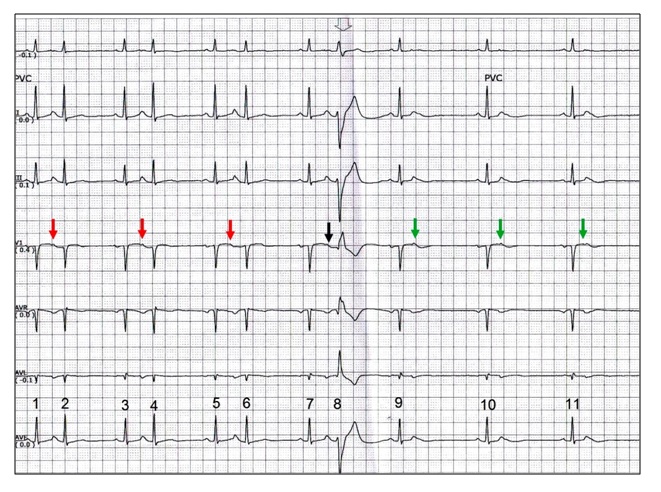 ECG Rhythms: Aberrancy
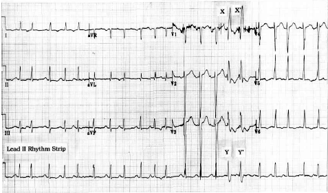 Multifocal Premature Ventricular Contractions
 Premature Ventricular Contractions (PVCs) ECG (Example 3), 50% OFF
%20Vs.%20Premature%20Atrial%20Contraction%20(Single)%20.webp) Premature Ventricular Contraction (PVC) Vs Premature Atrial
Premature Ventricular Contraction (PVC) Vs Premature Atrial
 A, Twelve‐lead ECG before the PVI of persistent AF and bigeminal PVCs
 Atrial Fibrillation and PVCs, How Do They Compare?Atrial Fibrillation
 ECG Learning Center - An introduction to clinical electrocardiography
 VT AF PVCs | ECG-Cafe
 The ECG and found peaks by the algorithm (a) of a patient with PVCs
 ECG Interpretation - 34 Coupled Pvc's | Ekg interpretation, Ecg
 VT AF PVCs | ECG-Cafe
 ECG Educator Blog : Premature Ventricular Contraction (PVC)
 Premature Ventricular Complex (PVC) • LITFL • ECG Library Diagnosis
 Premature Ventricular Contractions (PVCs) ECG Review | Learn the Heart

 Premature Ventricular Complex (PVC) • LITFL • ECG Library Diagnosis
 ECG: Premature Ventricular Complexes (PVC) - YouTube
 12-lead EKG showing frequent premature ventricular complexes (PVC
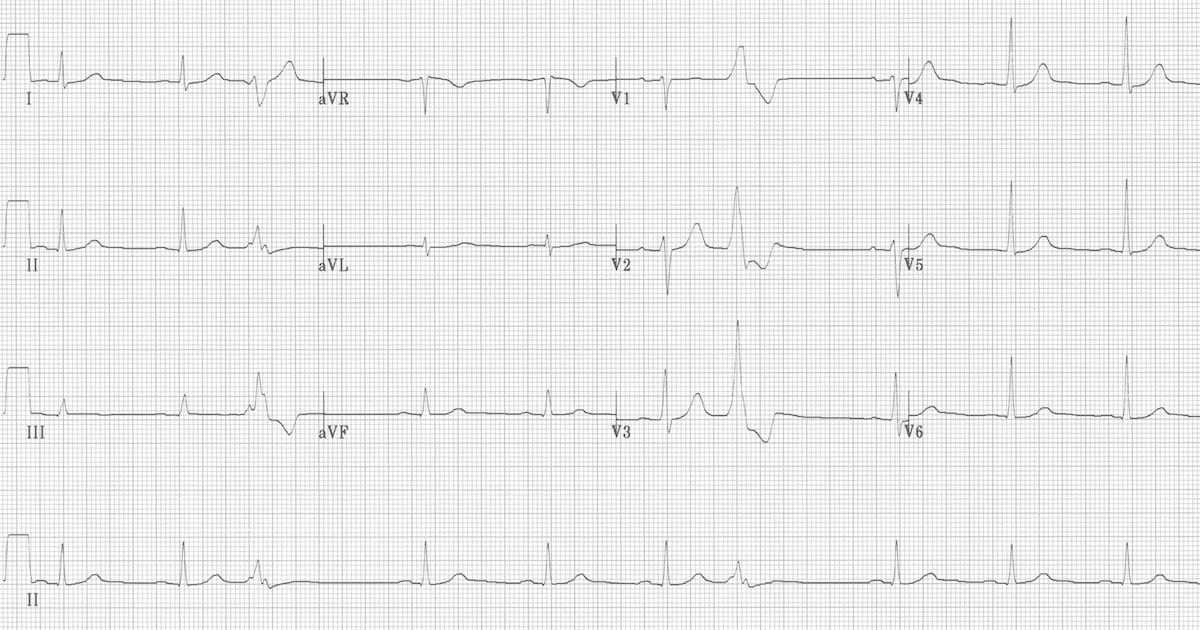 Premature Ventricular Complex (PVC) • LITFL • ECG Library Diagnosis
 EKG NOV 9 - NOV 13
 R on T Premature Ventricular Complexes (PVC) Simplified | ECGEDUcom

 ECG showed AF this morning for maybe 2 minutes and has been normal
