Left ventricular (LV) summit: boundaries, anatomic landmarks, typical ECG morphologies. LV summit the superior portion the LV (star, B) an important anatomic landmark it the region the epicardial surface, the left main coronary artery (LMCA) bifurcates is recognized the commonest source idiopathic .
 This region the highest portion the LV epicardium, the bifurcation the left main coronary artery (LMCA), accounts up 14.5% LV VAs. 2 complex relationships the left ventricular summit (LVS) surrounding structures underscore importance understanding anatomy this region the of .
 The image shows successful ablation an LVS PVC the LV subvalvular endocardium the LCC. A) image shows baseline ECG 2 PVCs. second PVC a clinical PVC (marked the arrow) an RBBB pattern, right inferior axis, a QS pattern lead I.
 Three months later, patient asymptomatic the 24-hour ambulatory ECG monitoring showed PVC burden 0.1% any antiarrhythmic agent. Figure 4: . performed stepwise catheter ablation the LV-summit PVC origin site adjacent severe coronary artery stenosis a 3D electroanatomic mapping system a single case .
 LV 32 (14.5%) (LV ostium 27 crux 5), mitral annulus 33 (14.9%), fascicles the left bundle-branch 40 (18.1%), papillary muscles 23 (10.4%), other sites 5 (2.3%). subjects the present study the 27 patients (13 men, 46 11 years, 24 70) a site the VA origin the epicardial LV
 Deductive Electrocardiographic Analysis of Left Ventricular Summit
 Muser Santangeli; Ablation LV Summit Communicating Vein VAs Circ Arrhythm Electrophysiol. 2018;11:e006105. DOI: 10.1161/CIRCEP.117.006105 January 2018 4 . institution, the decision map summit-CV discretional. ECG characteristics summit-CV arrhythmias reflected origin the posterior
 The PVC displayed QRS duration 148 ms, QS pattern lead I, maximum deflection index 0.8, intrinsicoid deflection time 52 ms, an aVL/aVR Q-wave ratio 1.6. features suggestive an epicardial origin the LV outflow tract (ie, LV summit).
 Background: early precordial electrocardiographic (ECG) characteristics useful differentiate left-sided the right-sided outflow tract ventricular arrhythmia (OTVA), patterns predict origin the septal margin the left ventricular (LV) summit. Objective: purpose this study to report mapping ablation characteristics a ECG pattern left .
 ABSTRACT. left ventricular (LV) summit the usual source epicardial idiopathic premature ventricular contractions (PVCs).A 56-year-old male patient presented the cardiology outpatient clinic palpitations dyspnea. Twelve-lead electrocardiography performed admission revealed monomorphic PVCs precordial QRS transition the V1 derivation an rS pattern the D1 .
 laboratory istheapproach toVAs arising fromthe summit the left ventricle (LV). region the highest portion the LV epicardium, the bifurcation the left main coronary artery (LMCA), accounts up 14.5% LV VAs.2 complex relationships the left ventricular summit (LVS) surrounding structures under-
 Ventricular Arrhythmias from the Left Ventricular Summit - Cardiac
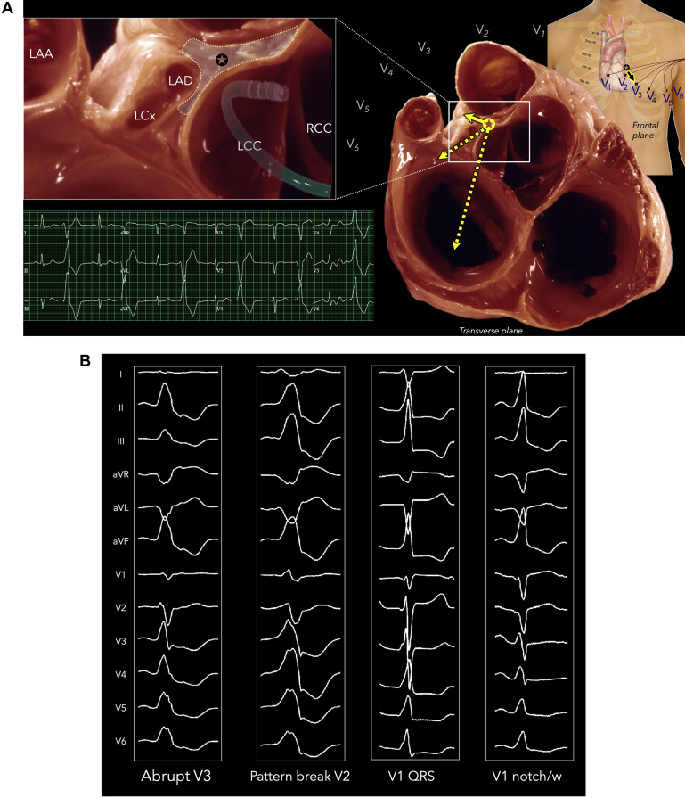 Left Ventricular Summit Arrhythmias with an Abrupt V3 Transition
 Bipolar Radiofrequency Catheter Ablation of Left Ventricular Summit
 Deductive Electrocardiographic Analysis of Left Ventricular Summit
 How to map and ablate left ventricular summit arrhythmias - Heart Rhythm
 Ablation strategies for intramural ventricular arrhythmias - Heart Rhythm
 Differentiating Right- and Left-Sided Outflow Tract Ventricular
 Novel technique targeting left ventricular summit premature ventricular
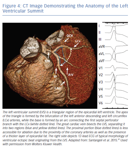 CT Image Demonstrating the Anatomy of the Left Ventricular Summit
 Left Ventricular Summit Arrhythmias with an Abrupt V3 Transition
 Fermin Carlos Garcia on Twitter: "ECG Pre ablation, no PVC post
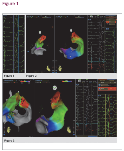 76/Successful catheter ablation of PVC from distal great cardiac vein
 Idiopathic Ventricular Arrhythmias Originating From the Left
 How to map and ablate left ventricular summit arrhythmias - Heart Rhythm
 Intramural Venous Ethanol Infusion For Refractory, 42% OFF
 | An example of successful ablation of premature ventricular complex
 Catheter Ablation of Arrhythmias Originating From the Left Ventricular
 Romero 12 11 20 Advanced Techniques for LV Summit PVCs - YouTube
 Deductive Electrocardiographic Analysis of Left Ventricular Summit
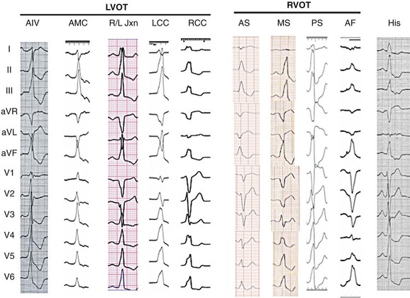 Outflow Tract Ventricular Tachyarrhythmias: Mechanisms, Clinical
 How to Ablate Ventricular Tachycardia from the Left Ventricular Summit
 LV summit venous anatomy On the left, schematic display of LV summit
 Catheter Ablation of Premature Ventricular Contractions From the Left
 Surface 12-lead ECG of the clinical tachycardia (left) suggestive of a
 (PDF) A Comprehensive Review of Left Ventricular Summit Ventricular
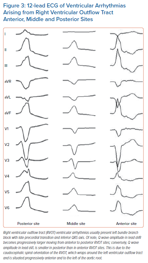 12-lead ECG of Ventricular Arrhythmias Arising from Right Ventricular
 Anatomy for Ventricular Tachycardia Ablation in Structural Heart
 Ventricular Arrhythmias | Thoracic Key

