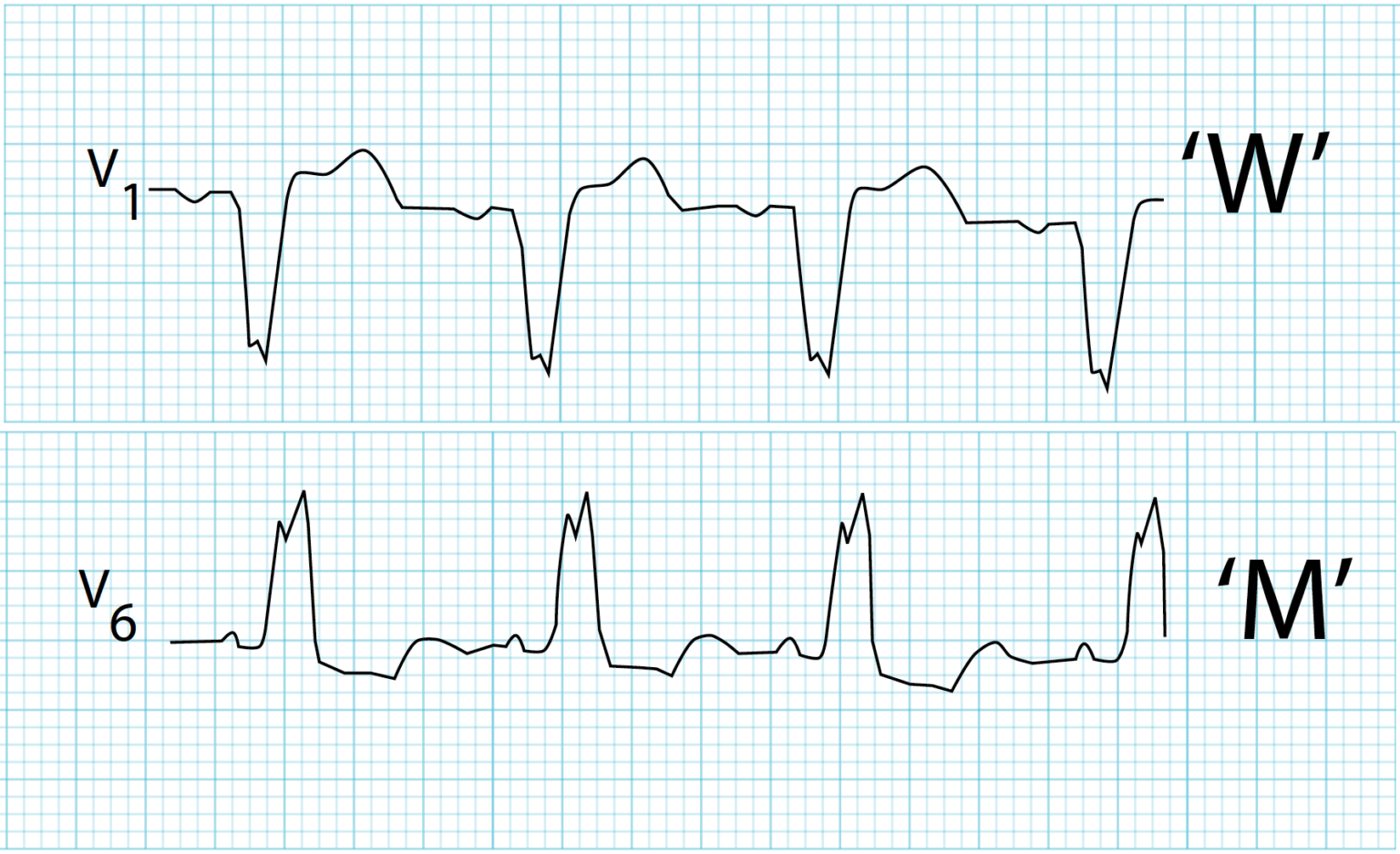
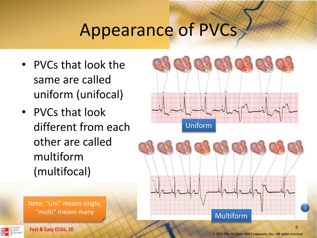
Unifocal — arising a single ectopic focus; PVC identical; Multifocal — arising two more ectopic foci; multiple QRS morphologies; origin each PVC be discerned the QRS morphology: PVCs arising the ventricle a left bundle branch block morphology (dominant wave V1)
 A: Left bundle branch pacing (LBBP) nonischemic cardiomyopathy (NICM) left bundle branch block (LBBB). (i) Premature ventricular contractions (PVCs) with changing morphology QS qR pattern (PVC1) lead 1 noted rapid rotation. (ii) Left bundle branch (LBB) paced QRS mimicked PVC1 duration 124 ms peak left ventricular activation time (pLVAT) 78ms.
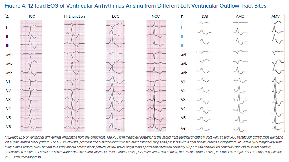
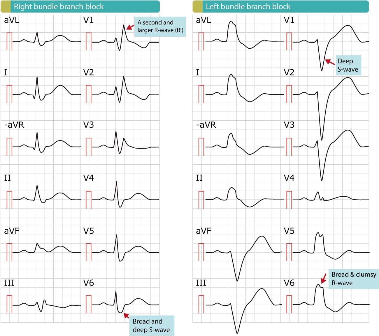
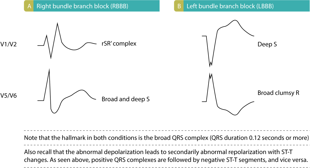
 The QRS morphology EKG predict PVCs site origin. a broad general rule, right ventricular ectopic pacemaker generates ventricular complex left bundle branch block (LBBB) pattern, the left ventricular ectopic pacemaker generates ventricular complex right bundle branch block (RBBB) pattern 2. right left ventricular outflow tracts aortic cusp .
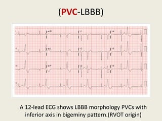 The morphology PVC lead V1 provide clues PVC site origin. PVC has morphology RBBB lead V1 a site origin the left ventricle, PVC with LBBB morphology lead V1 usually sourced the ventricle. not absolute, algorithms determining PVC site origin by .
 PVC morphology LBBB with very late transition superior axis. Figure 1: (A) Typical RVOT PVCs a distance runner, a LBBB pattern, precordial transition V4, inferior axis. (B) ventricular apical PVCs a patient early ARVC. PVC morphology LBBB with very late transition superior axis.
 Premature ventricular complexes (PVCs) extremely common, in majority individuals undergoing long-term ambulatory monitoring. Increasing age, taller height, higher blood pressure, history heart disease, performance less physical activity, smoking predict greater PVC frequency. the fundamental of PVCs remain largely unknown, potential .
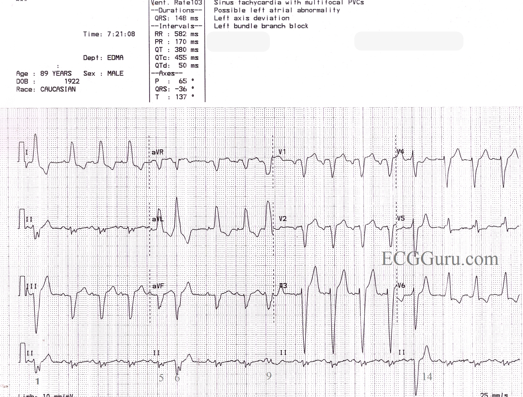
 Left bundle branch block morphology most common type 'benign PVCs' originates the RVOT is characterized a LBBB with inferior axis morphology (infundibular pattern). this context, LBBB pattern defined a negative QRS complex lead V1, negative QRS complex lead aVL, a positive QRS the inferior .
 Much research premature ventricular complexes (PVCs) used inferior axis left bundle block pattern (I-LBBB) a unique category PVCs, with assumption PVCs with pattern originate the outflow tract (OT) region are idiopathic.1,2 However, is established these assumptions not true all I-LBBB PVCs.3 distinction OT non-OT PVCs .
 Premature ventricular contractions left bundle branch block pattern (PVC‐LBBB) seen 41% the children (Figure 1), with right bundle branch block pattern (PVC‐RBBB) seen 36% (Figure 2), the morphology undetermined 23%. was observed although mean percentage PVC‐LBBB not change, PVC .
 Outflow tract ventricular arrhythmias (OTVAs) the common type idiopathic VA. typically presents young patients—and a notably increasing incidence. 1 is classically benign, focal arrhythmia patients be highly symptomatic refractory medical therapy. Moreover, frequent ectopy progress a premature ventricular complex (PVC)-induced cardiomyopathy.
 Role of His Refractory Premature Ventricular Complexes in the
 | A patient who suffered from premature ventricular contraction (PVC
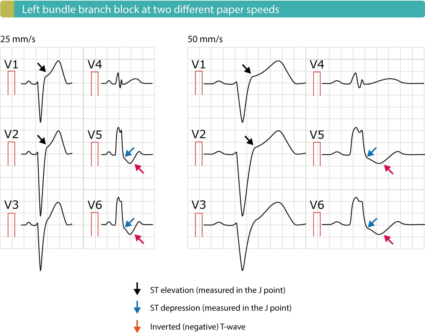 Left bundle branch block (LBBB): ECG criteria, causes, management - The
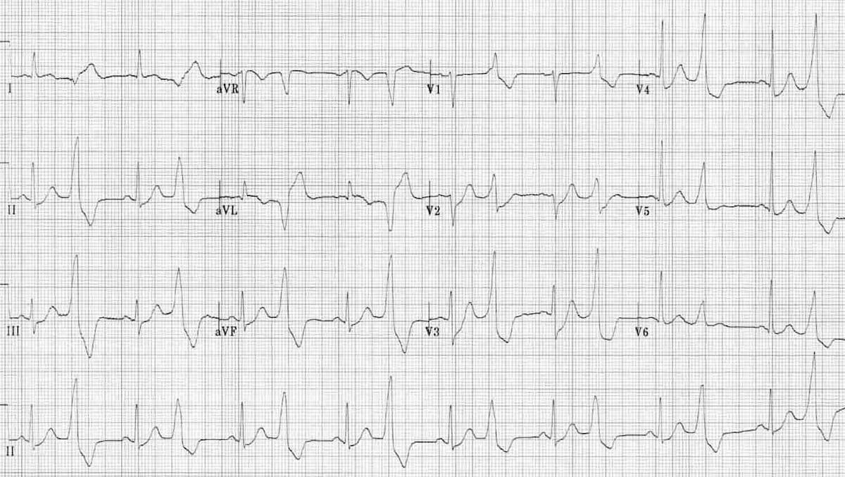 Premature Ventricular Complex (PVC) • LITFL • ECG Library Diagnosis
 Electrocardiogram (ECG) Diagnosis Of Ventricular Arrhythmias
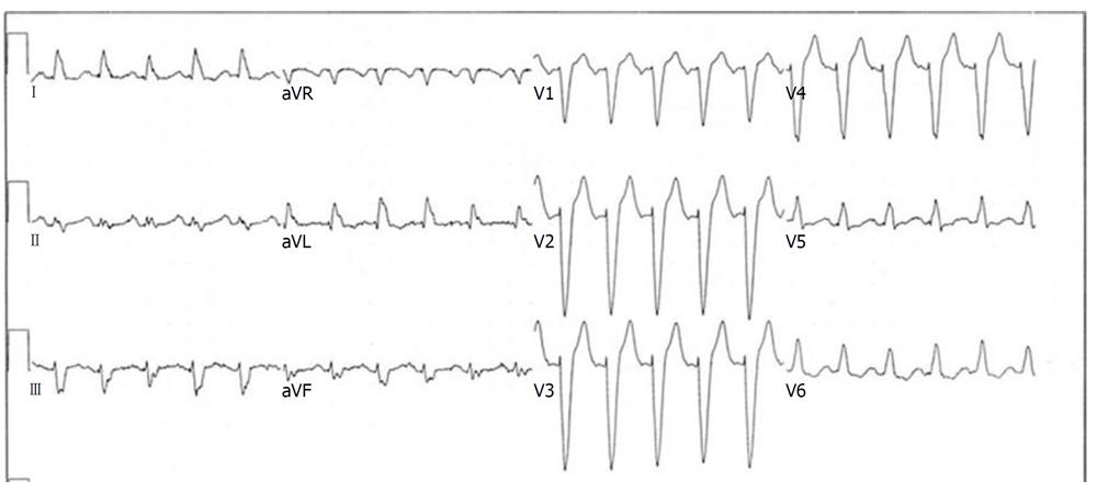 Differential diagnosis of tachycardia with a typical left bundle branch
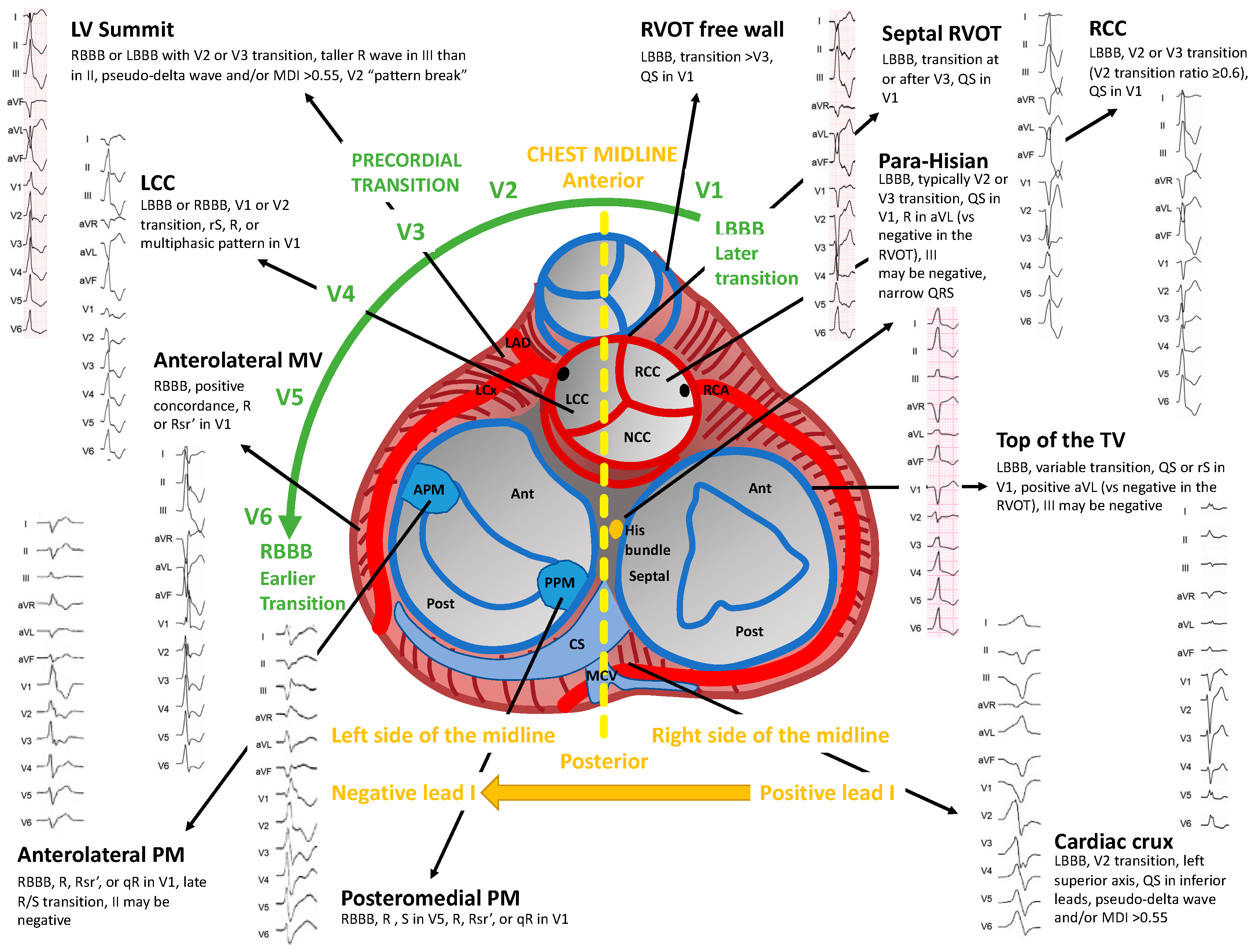 Diagnostics | Free Full-Text | Diagnosis and Treatment of Idiopathic
 Figure1 ECG of PVC1 and PVC2 showed as LBBB morphology and RBBB
 ECG Rhythms: Aberrancy
 ECG after resuscitation exhibiting ST-segment elevation in V1 and V2
 Premature Ventricular Complex (PVC) • LITFL • ECG Library Diagnosis

 Clinical characteristics of index patient and LMNA mutation-positive
 Left Bundle Branch Block (LBBB) • LITFL • ECG Library Diagnosis
 Evaluation and Management of Premature Ventricular Complexes | Circulation
 Left Bundle Branch Block (LBBB) • LITFL • ECG Library Diagnosis
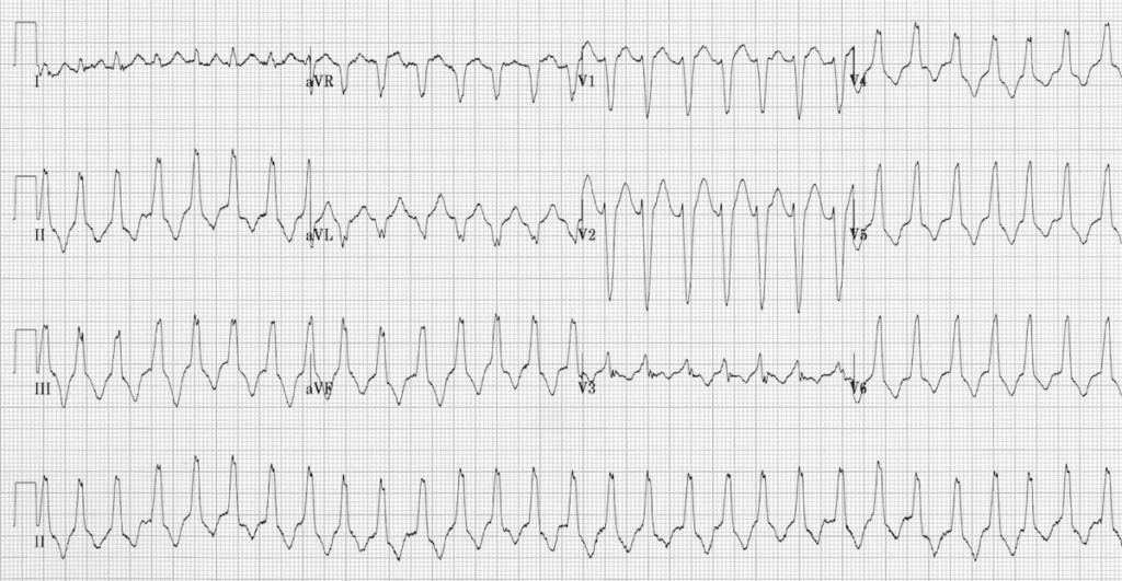 Arrhythmogenic Right Ventricular Cardiomyopathy (ARVC) • ECG Library
 (A) Clinical VT of LBBB morphology was inducible by programmed
 Differentiating Right- and Left-Sided Outflow Tract Ventricular
 Left Bundle Branch Block (LBBB) • LITFL • ECG Library Diagnosis
 The difference between the normal narrow QRS and wide QRS as seen in
 (A) Morphology of the second PVC (B) Earliest activation is seen under
 Left Bundle Branch Block (LBBB) • LITFL • ECG Library Diagnosis
 Right bundle branch block (RBBB): ECG, criteria, definitions, causes
 Premature Ventricular Complex (PVC) • LITFL • ECG Library Diagnosis
 PPT - Ventricular Dysrhythmias PowerPoint Presentation, free download
 Intraventricular conduction delay: bundle branch blocks & fascicular
 How to Ablate Non-Outflow Right Ventricular Tachycardia | Thoracic Key
 Left bundle branch block - WikEM
